Datei:Hepatocellular carcinoma 1.jpg
Hepatocellular_carcinoma_1.jpg (550 × 368 Pixel, Dateigröße: 38 KB, MIME-Typ: image/jpeg)
![]()
Diese Datei und die Informationen unter dem roten Trennstrich werden aus dem zentralen Medienarchiv Wikimedia Commons eingebunden.
![]()
Beschreibung
| Beschreibung |
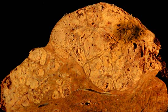
Hepatocellular carcinoma This specimen is from a 50ish woman who presented to the hospital with abdominal pain and ascites. The radiologist recovered what appeared to be whole blood on paracentesis. Cytological exam of the bloody fluid showed no evidence of malignancy. Liver function tests were abnormal, and serologic tests were positive for antibody to hepatitis C. The patient deteriorated rapidly and died within a few days. The autopsy showed this hepatocellular carcinoma occupying much of the volume of a cirrhotic liver. Furthermore, the tumor had invaded the diaphragm and ruptured into the peritoneal cavity, causing the bloody ascites. The photo shows a view of a longitudinal slice taken through the full length of the liver. The photos were shot with a Minolta X-370 with 100 mm bellows lens on Kodak Elite ISO 100 transparency film. The specimen was sliced fresh and fixed in formalin overnight, then briefly immersed in 70% alcohol to retrieve some of the native color and dull the surface reflections. Photograph by Ed Uthman, MD. Public domain. Posted 23 Sep 00 |
| Quelle | http://web2.airmail.net/uthman/specimens/index.html |
| Urheber | |
| Genehmigung (Weiternutzung dieser Datei) |
PD |
Lizenz
| Dieses Werk wurde von seinem Urheber Ed Uthman als gemeinfrei veröffentlicht. Dies gilt weltweit. In manchen Staaten könnte dies rechtlich nicht möglich sein. Sofern dies der Fall ist: Ed Uthman gewährt jedem das bedingungslose Recht, dieses Werk für jedweden Zweck zu nutzen, es sei denn, Bedingungen sind gesetzlich erforderlich.
|
Dateiversionen
Klicke auf einen Zeitpunkt, um diese Version zu laden.
| Version vom | Vorschaubild | Maße | Benutzer | Kommentar | |
|---|---|---|---|---|---|
| aktuell | 12:14, 5. Jun. 2006 | | 550 × 368 (38 KB) | Patho | {{Information| |Description=Hepatocellular carcinoma This specimen is from a 50ish woman who presented to the hospital with abdominal pain and ascites. The radiologist recovered what appeared to be whole blood on paracentesis. Cytological exam of the blo |
Dateiverwendung
Die folgende Seite verwendet diese Datei:
Globale Dateiverwendung
Die nachfolgenden anderen Wikis verwenden diese Datei:
- Verwendung auf ar.wikipedia.org
- Verwendung auf ast.wikipedia.org
- Verwendung auf az.wikipedia.org
- Verwendung auf be.wikipedia.org
- Verwendung auf bs.wikipedia.org
- Verwendung auf ca.wikipedia.org
- Verwendung auf cs.wikipedia.org
- Verwendung auf de.wikibooks.org
- Verwendung auf el.wikipedia.org
- Verwendung auf en.wikipedia.org
- Hepatocellular carcinoma
- Alcohol and cancer
- Portal:Medicine/Selected article/50, 2007
- Portal:Medicine/Selected Article Archive (2007)
- Obesity-associated morbidity
- Cirrhosis
- Portal:Viruses
- Portal:Viruses/Selected article
- Portal:Viruses/Selected article/10
- User:Daniel Mietchen/Wikidata lists/Items with Disease Ontology IDs
- Verwendung auf eo.wikipedia.org
- Verwendung auf es.wikipedia.org
- Verwendung auf eu.wikipedia.org
- Verwendung auf fa.wikipedia.org
- Verwendung auf fi.wikipedia.org
- Verwendung auf fr.wikipedia.org
- Verwendung auf gl.wikipedia.org
- Verwendung auf he.wikipedia.org
- Verwendung auf hi.wikipedia.org
- Verwendung auf hy.wikipedia.org
- Verwendung auf id.wikipedia.org
- Verwendung auf it.wikipedia.org
- Verwendung auf ja.wikipedia.org
Weitere globale Verwendungen dieser Datei anschauen.